
Light Microscope
Light microscopes use glass lenses and visible light to magnify microbes, cells, and tiny organisms up to 1,000 times.
- You can view live specimens and use stains to highlight details.
- Staining Techniques:
- Gram Staining helps identify bacteria as Gram-positive (purple) or Gram-negative (pink), essential for choosing antibiotics.
- Fluorescent Stains: Glow under UV light to detect bacteria and viruses.
It is limited in magnification, so it cannot see viruses or tiny cell structures.
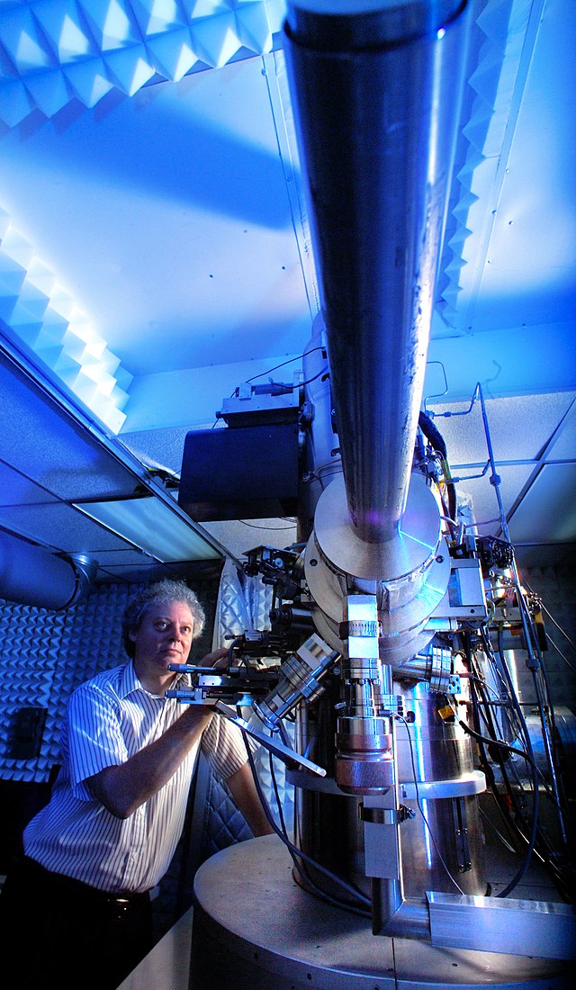
Electron Microscope
Electron microscopes use beams of electrons to magnify objects up to 10 million times, revealing viruses, cell structures, and molecules that are too small for light microscopes.
- Since electrons cannot pass through air or glass, samples must be placed in a vacuum.
- Electron microscopes use special stains made of heavy metals (e.g., lead, osmium, uranium) to improve contrast.
Two main types:
- Scanning Electron Microscope (SEM): Creates 3D images of surfaces.
- Transmission Electron Microscope (TEM): Shows internal cell structures in fine detail.
Applications in Microbiology
Microscopes are crucial in microbiology, helping scientists study, diagnose, and innovate in various fields.
Disease Diagnosis: Identifying bacteria, viruses, and fungi in medical labs to detect infections.
Antibiotic Research: Studying bacterial structures to develop effective antibiotics and treatments.
Forensic Science: Analyzing microbial evidence at crime scenes for investigations.
Environmental Studies: Examining microbes in soil and water to monitor ecosystems and pollution.
Biotechnology & Vaccines: Developing new medicines, vaccines, and bioengineered microbes for health and industry.
Fun Facts
Here are some interesting facts about microscopes:
- Antonie van Leeuwenhoek first saw bacteria and protists using a simple microscope and called them “animalcules.”
- Louis Pasteur and Robert Koch used microscopes to show that bacteria cause diseases, leading to vaccines and sterilization methods.
- Microscopes revealed the double-helix structure of DNA, thanks to electron microscopy.
- Electron microscopes allowed scientists to see viruses for the first time, leading to advancements in vaccine development.
Review
Let’s quickly recap what we learned about microscopes:
- What type of staining does a light microscope use? Gram Staining & Fluorescent Stains
- Where must the samples be placed in an electron microscope? Vacuum
- Which microscope provides a 3D image of a microbe’s surface? Scanning Electron Microscope
- What type of stains does an electron microscope use? Heavy Metals

Recent Comments